Abstract
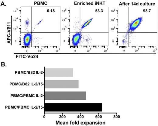
Invariant natural killer T (iNKT) cells have been shown to play an important role in immune regulation, defense against pathogens, and cancer immunity, despite representing a minority population of lymphocytes. In particular, emerging murine and human data demonstrate that within this immunoregulatory capacity, iNKT cells function to suppress graft-versus-host disease without loss of the graft-versus-tumor effect, making them ideal to exploit in hematopoietic cell transplantation. Given the key functions of this relatively understudied population, the ability to ex vivo expand human iNKT cells has vital scientific and clinical applications. Therefore, we sought to determine the optimal conditions for iNKT cell expansion and the effects of expansion method on the iNKT cell cytokine production profile. Several expansion culture conditions were explored following initial magnetic bead-based iNKT enrichment and stimulation with α-galactosylceramide (α-GalCer)-loaded irradiated autologous peripheral blood mononuclear cells (PBMCs) on day 0: inclusion of either IL-2 or IL-2 plus IL-15 and day 10 restimulation with either α-GalCer-loaded irradiated autologous PBMCs or α-GalCer-loaded irradiated K562 cells engineered to express CD1d, CD86, 4-1BBL, and OX40L ("B82" cells; Tian G et al. J Clin Invest. 2016 Jun 1;126(6):2341-55). We found significant heterogeneity in the numbers of circulating iNKT cells in healthy donors, ranging from 0.05-0.3% of CD3+ cells, as well as in the fold expansion obtained following two weeks of in vitro culture. Ex vivo expansion using B82 cells for day 10 stimulation and IL-2 alone routinely yielded viability >90% and purity >95% (Figure 1A), and we obtained a median of 3.5x10⁷ iNKT cells starting from a median of 7.2x10⁸ PBMCs following two weeks of expansion. However, we found that when iNKT cells were expanded with autologous PBMCs used on day 10 for restimulation, fold expansion was higher than when B82 cells were used on day 10 and the inclusion of IL-15 along with IL-2 in the culture media led to substantially enhanced expansion (mean fold expansion for PBMC with IL-2: 458; B82 with IL-2: 314; PBMC with IL-2 plus IL-15: 634; B82 with IL-2 plus IL-15: 375; Figure 1B). Following PMA/ionomycin stimulation, cytokine production profiles were similar regardless of expansion conditions, with nearly all iNKT cells producing IFNγ (ranging 94.4-99.9%), and a smaller fraction of iNKT cells producing IL-4 (ranging 21.0-36.1%). These data describe the ex vivo expansion capacity of iNKT cells and culture conditions which lead to optimal expansion. These results contribute to our understanding of human iNKT cells and could inform the design of future clinical trials utilizing iNKT cells.
No relevant conflicts of interest to declare.
Author notes
Asterisk with author names denotes non-ASH members.


This feature is available to Subscribers Only
Sign In or Create an Account Close Modal